The Imaging Services Department at Tomah Health serves all patient age groups and offers several radiological examinations and procedures. The procedures and treatments act as a diagnostic tool that assists the patient’s health care provider in the determination of diagnosis and treatment options.
Imaging Services
Diagnostic radiology is a field of medicine in which non-invasive imaging is utilized to allow visualization of body tissues, organs, and bones to diagnose and treat patients. State of the art equipment provides lower doses of radiation and high detailed images for interpretation. Fluoroscopy is a type of medical imaging that shows a continuous X-ray image on a monitor, much like an X-ray movie. During a fluoroscopy procedure, an X-ray beam is passed through the body. The image is transmitted to a monitor so the movement of a body part or of an instrument or contrast agent (“X-ray dye”) through the body can be seen in detail.
An echocardiogram (echo) utilizes high frequency sound waves (ultrasound) to produce images of the heart chambers, valves, walls, and vessels that are attached to the heart. The ultrasound scan of the heart creates moving pictures displaying the structure and functions of the heart for diagnosis and monitoring of heart disease. An echo can identify leaky or tight heart valves.
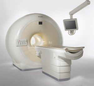
Magnetic resonance imaging is a non-invasive and painless imaging technique that uses strong magnetic fields, magnetic field gradients, and radio waves to generate detailed images of the organs and tissues within the body. Unlike CT and Xray, MRI does not utilize radiation. Our state of art scanner has a 70cm bore (wide tube) to help ease patient anxieties. Blankets and cushions are provided by technologists to ensure patients are comfortable as possible.
Nuclear medicine exams use very small amounts of radioactive materials, called radiopharmaceuticals or tracers, to monitor, diagnose and treat medical conditions within the body. These images show the function, size and shape of an organ, tissue, bone or body system.
Computed tomography (CT) is a diagnostic imaging test that creates detailed images of internal organs, bones, soft tissue, and blood vessels that cannot be seen in conventional xray. Cross sectional images are generated during the scan which allow for images to be reformatted into three-dimensions. CT is a fast, painless, and noninvasive scan that is often the best in detecting many different cancers. This includes the detection of tumors, along with the size and location. In emergencies, CT can reveal internal injuries and bleeding quick enough to help save lives.
Ultrasound is a type of imaging that uses high-frequency sound waves to look at organs and structures inside the body. Ultrasound is safe, noninvasive, and does not utilize ionizing radiation. The pictures produced from ultrasound help diagnose the causes of pain, swelling, infections, and helps to examine a baby in pregnant women.
Tomah Health has been awarded a three-year term of accreditation in computed tomography (CT) by the American College of Radiology (ACR). The ACR gold seal of accreditation represents the highest level of image quality and patient safety.
Our Team
The department is staffed every day by technologists who perform various radiological procedures. Those on staff include:
- Imaging Director & Coordinator
- Registered Imaging Technologists
- Registered Sonographer
- Registered CT Technologists
- Registered MRI Technologists
The team of qualified staff maintain a level of performance that contributes to high quality imaging and efficiencies with all services provided. A team of Radiologists provide professional coverage for consultation and treatment of patients.
For more information on the services provided by our Imaging Services Department, please call (608) 377-8247.